Analysis of main performance parameters of scanning electron microscope
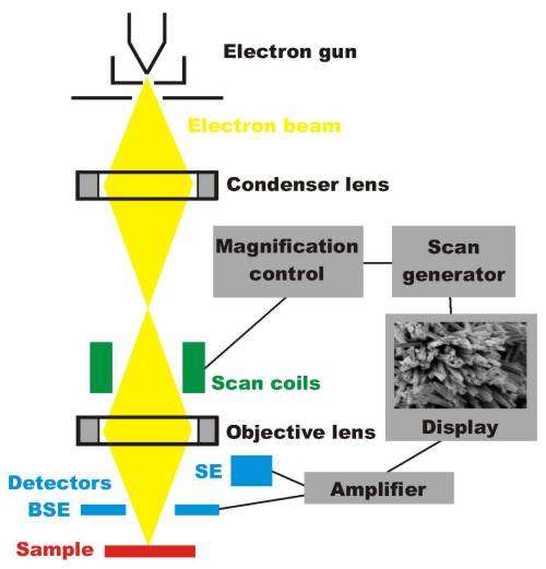
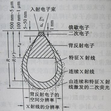
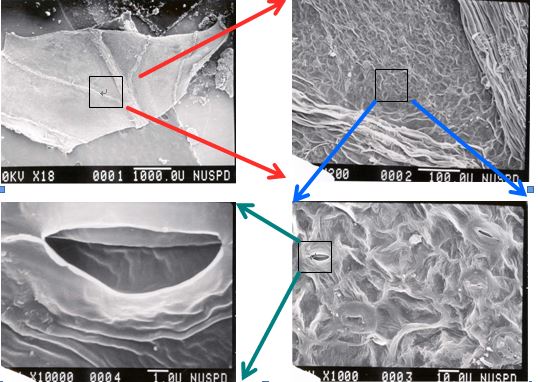
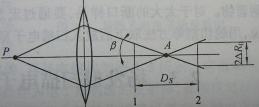
This article introduces the main parameters of SEM: resolution, magnification, depth of field. Resolution Resolution is the most important performance indicator of SEM. For imaging, it refers to the minimum distance between two points. For micro-area component analysis, it refers to the smallest area that can be analyzed. The resolution of a scanning electron microscope is determined by measuring the minimum distance between two particles (or regions) in an image by dividing the minimum spacing measured on the image by the magnification at a known magnification. The resulting value is the resolution. The resolution of the scanning electron microscope is related to the type of detection signal. The following table shows the imaging resolution of the main signal of the scanning electron microscope. Signal backscattered electron secondary electron characteristic X-ray absorption electron Auger electron Resolution / nm50-2005-10100-1000100-10005-10 It can be seen from the data in the table that the resolution of secondary electrons and Auger electrons is not high, and the resolution of characteristic X-ray modulation into microscopic images is the lowest. The reason why the difference between the resolutions caused by different signals can be explained by the following figure, the electron beam entering the surface of the light element sample will cause a drop volume. The incident electron beam will move in this volume before it is absorbed or scattered by the sample. Secondary electrons and Auger electrons can only escape in the shallow surface of the sample because of their low energy and short mean free path. Under normal circumstances, the surface thickness of the sample of Auger electrons is about 5- 2 nm, the layer depth of the excited secondary electrons is in the range of 5-10 nm. When the incident electron beam enters the shallow surface, it has not expanded laterally. Therefore, the secondary electron and the Auger electron can only be excited in a cylinder with a diameter corresponding to the incident electron beam spot, because the beam spot diameter is an image. The size of the detection unit (image point), so the resolution of these two electrons is equivalent to the diameter of the beam spot. When the incident electron beam enters the deep part of the sample, the range of lateral expansion becomes larger, and the backscattered electron energy excited from this range is high, and they can eject the surface from the deep part of the sample, and the effect of lateral expansion The size is the imaging unit of backscattered electrons, which greatly reduces its resolution. The incident electron beam can also excite characteristic X-rays in deeper parts of the sample. From the perspective of the X-ray volume, the resolution is lower than that of backscattered electrons. It should be noted that when the electron beam is injected into the heavy element sample, the action volume does not appear as a drop, but is hemispherical. The electron beam expands laterally as it enters the surface, so when analyzing heavy elements, even if the beam spot of the electron beam is very small, a higher resolution cannot be achieved. The resolution of a scanning electron microscope depends on the diameter of the incident electron beam. The smaller the diameter, the higher the resolution, but the resolution is not directly equal to the incident electron beam diameter. Because the effective excitation range of the electron beam in the sample greatly exceeds the incident electron beam diameter. In addition, the resolution of the scanning electron microscope is affected by factors such as the atomic number of the sample, the stray magnetic field, and the mechanical vibration, in addition to the influence of the electron beam diameter and the type of the modulated signal. Magnification When the incident electron beam is raster scanned, if the amplitude of the electron beam scanning on the surface of the sample is As, and the amplitude of the synchronous scanning of the cathode ray on the illuminating screen is Ac, the magnification may be expressed as M=Ac/As. Since the size of the screen is constant, the change in magnification is achieved by changing the scanning amplitude As of the electron beam on the surface of the sample. At present, the magnification of the commercial scanning electron microscope can be continuously adjusted from 20 times to 200,000 times. Depth of Field Depth of field refers to the range of ability of a lens to simultaneously focus an image on various parts of a rugged sample. This range is expressed by a distance. If the depth of field is Ds, as long as the surface height of the sample is less than Ds, the surface topography of the sample can be clearly reflected on the screen. It is precisely because of the large depth of field of the scanning electron microscope that it is especially suitable for the analysis and observation of rough surfaces and fractures; the image is full of three-dimensionality, realism, easy to identify and explain. In our kitchen storage product page, you can find salad bowl, seasoning jars, sauce bottle, glass storage jars and plate rack. Salad Bowl,Seasoning Jars,Glass Storage Jars,Plate Rack,Sauce Bottle Shangwey Cookware Co.,Ltd of Jiangmen City , https://www.shangwey.com